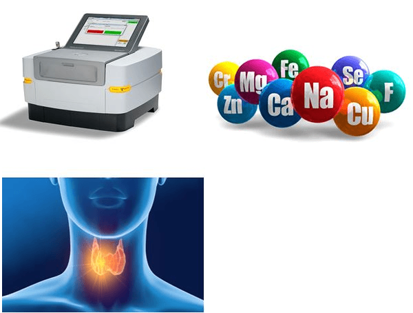
DIAGNOSIS OF THYROID MALIGNANCY USING TRACE ELEMENTS OF NODULAR TISSUE DETERMINED BY X-RAY FLUORESCENCE ANALYSIS
Keywords:
Diagnosis of thyroid malignancy, Normal thyroid, Thyroid nodules, Trace elements, Energy-dispersive X-ray fluorescent analysisAbstract
Background: Benign (TBN) and malignant (TMN) thyroid nodules are a common thyroid lesion. The differentiation of TMN often remains a clinical challenge and further improvements of TMN diagnostic accuracy are warranted. The aim of this study is to evaluate the possibilities of using differences in trace element contents (TEs) in nodular tissue to diagnose thyroid malignancies and to determine the sensitivity, specificity, and accuracy for most informative TEs in the diagnosis of TMN.
Methods: Contents of TEs such as bromine (Br), copper (Cu), iron (Fe), iodine (I), rubidium (Rb), strontium (Sr), and zinc (Zn) were prospectively evaluated in “normal” thyroid (NT) of 105 individuals as well as in nodular tissue of thyroids with TBN (79 patients) and to TMN (41 patients). Measurements were performed using energy-dispersive X-ray fluorescent analysis.
Results: It was observed that in TMN tissue the mean mass fractions of I and Zn were lower while the mean mass fraction of Rb was higher than in NT and TBN tissue. It was demonstrated that the I contents is nodular tissue is the most informative parameter for the diagnosis of thyroid malignancy. It was found that “Sensitivity”, “Specificity” and “Accuracy” of TMN identification using the I level in the needle biopsy of affected thyroid tissue (87±5%, 96±2% and 94±2% respectively) were significantly higher than that made using ultrasound screening and cytological test of fine needle aspiration biopsy.
Conclusions: It was concluded that determination the I level in a needle biopsy of TNs using energy-dispersive X-ray fluorescent analysis, is a fast, reliable, and informative diagnostic tool that can be successfully used as an additional test of thyroid malignancy identification.

Peer Review History:
Received: 4 February 2022; Revised: 9 March; Accepted: 28 April; Available online: 15 May 2022
Academic Editor: Dr. Emmanuel O. Olorunsola , Department of Pharmaceutics & Pharmaceutical Technology, University of Uyo, Nigeria, olorunsolaeo@yahoo.com
Reviewers:
 Prof. Dr. Hassan A.H. Al-Shamahy, Sana'a University, Yemen, shmahe@yemen.net.ye
 Dr. U. S. Mahadeva Rao, Universiti Sultan Zainal Abidin, Terengganu Malaysia, raousm@gmail.com
Downloads

Published
How to Cite
Issue
Section

This work is licensed under a Creative Commons Attribution-NonCommercial 4.0 International License.


 .
.