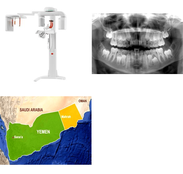
HISTOLOGIC AND RADIOGRAPHIC STUDY OF PATHOLOGIC CHANGE IN COMPLETE IMPACTED THIRD MOLARS DENTAL FOLLICLES
Keywords:
Dental follicle (DF), histopathological changes (HC), impacted third molars, oral pathology, radiographic width, Sana’a, YemenAbstract
Background: Prophylactic extraction of the asymptomatic impacted third molar is routinely practiced in Europe and the United States. The justification for prophylactic extraction includes the need to reduce the risk of pathologic changes such as cysts and tumors.
Objectives: This study aimed to study the histological and radiological changes in the tooth follicles of upper and lower complete impacted 3rd molars -which appeared radiologically normal.
Material and method: A prospective study included fifty patients aged 20 years and above who were referred to the Oral Surgery Clinic, Faculty of Dentistry, University of Sana'a. Patients had follicular space between (2.5mm -3mm) as measured by the panoramic X-ray. These teeth were removed surgically and the follicle was sent for histopathological examination.
Results: Most histopathological changes were in dental follicles with a size of <2.5 mm (86%), and only 14% with 2.5 mm - 3 mm. There was statistical significance between the smallest size of dental follicles with the incidence of pathological histological changes indicating a high probability of developing neoplasm (p =0.008). Of the 50 follicular patients, 28% showed HC, nine (64%) had ameloblastoma, four (29%) had a dentigerous cyst, and only one case (7%) had a multicalcified focus with islands of odontogenic epithelium. While 72% of the samples had normal follicles and non-specific chronic inflammatory cells. There is an association between female sex and pathological histological changes (12 females: 2 males, p =0.008), age group 21-25 years (93% HC), with mandibles (65% HC). Regarding angle and histopathological changes, 36% were vertical, 29% mesioangular, 14.2% horizontal and destioangular, and 7.1% buccoangular.
Conclusion: High incidence of HC occurred in patients with DF, and it was associated with smaller dental follicle size, most HC was ameloblastoma, followed by dentigerous cyst, while 72% of samples had normal follicles and non-specific chronic inflammatory cells. There is a correlation between female gender, younger age group, and jaw position with HC. Prophylactic extraction of the asymptomatic impacted third molar should be routinely practiced in Yemen, to reduce the risk of pathological changes, especially in females and younger age groups.

Peer Review History:
Received 2 December 2020; Revised 7 January 2021; Accepted 12 February; Available online 15 March 2021
Academic Editor: Rola Jadallah , Arab American University, Palestine, rola@aauj.edu
Reviewer(s) detail:
Dr. A.A. Mgbahurike , University of Port Harcourt, Nigeria, amaka_mgbahurike@yahoo.com
Dr. Alfonso Alexander Aguileral , University of Veracruz, Mexico, aalexander_2000@yahoo.com
Downloads

Published
How to Cite
Issue
Section

This work is licensed under a Creative Commons Attribution-NonCommercial 4.0 International License.


 .
.