
THREE-DIMENSIONAL EVALUATION OF THE FIRST CERVICAL VERTEBRAL MORPHOLOGY IN SKELETAL CLASS I AND III MALOCCLUSIONS IN YEMENI PATIENTS
-
 Etehad Al-Ghola
Orthodontics, Pedodontics and Prevention Department Faculty of Dentistry, Sana'a University, Yemen.
Etehad Al-Ghola
Orthodontics, Pedodontics and Prevention Department Faculty of Dentistry, Sana'a University, Yemen.
-
Ghamdan Al-Harazi
Orthodontics, Pedodontics and Prevention Department Faculty of Dentistry, Sana'a University, Yemen.
-
Hassan Abdulwahab Al-Shamahy
Departement of Basic Sciences, Faculty of Dentistry, Sana’a University, Republic of Yemen. Medical Microbiology and Clinical Immunology Department, Faculty of Medicine and Health Sciences, Sana’a University, Republic of Yemen.
Keywords:
Cervical vertebrae, Cone-beam computed tomography (CBCT), orthodontics, YemenAbstract
Introduction: The shape of the cervical vertebrae is known to be of great importance in the field of orthodontics, as it can be used to assess skeletal maturity. This study aimed to evaluate the shape of the cervical vertebrae in individuals with class I and III skeletal malocclusion.
Methods: The research data were collected from the orthodontic patient records by analyzing cone beam computed tomography of the selected individuals before treatment (n=52) this study was conducted between January 2022 and January 2023. The individuals were divided into two groups based on the ANB angle: class I group (n=26, male=12, female=14) and class III group (n=26, male=9, female=17). The ages ranged from 18 to 30 years, and they were all Yemeni.
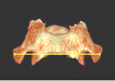
Results: The shape of the cervical vertebrae differs among individuals with different anteroposterior skeletal patterns. Cone beam radiographs of 52 patients were evaluated to assess the morphology of the first cervical vertebra in both Class I and Class III. Eight linear variables and one angular variable were compared in both groups, and there were significant differences between HOTDC1, LOAPC1, dorsal arch, HOTDC1, H1APC1, FOTDC1, and superior surface among individuals with different anteroposterior skeletal patterns.
Conclusion: The morphology of the cervical vertebrae was found to be influenced by the anteroposterior relationship of the maxilla to the mandible.
Peer Review History:
Received 11 December 2024; Reviewed 6 January 2025; Accepted 21 February; Available online 15 March 2025
Academic Editor: Dr. Ali Abdullah Al-yahawi , Al-Razi university, Department of Pharmacy, Yemen, [email protected]
Reviewers:
Dr. Naglaa Mohamed Ahmed Abd Elaal, Helwan University, Egypt, [email protected]
Dr. Nazim Hussain, North East Frontier Technical University, Arunachal pradesh, India, [email protected]
Downloads


Published
How to Cite
Issue
Section

This work is licensed under a Creative Commons Attribution-NonCommercial 4.0 International License.


 .
.