
PREVALENCE OF ORAL REACTIVE HYPERPLASTIC LESIONS AND ASSOCIATED RISK FACTORS IN A SAMPLE OF YEMENI DENTAL PATIENTS IN SEVERAL UNIVERSITIES AND PUBLIC HOSPITALS
-
 Shaima’a Abdulraqeeb Yahya Rajeh
Department of Basic Sciences, Faculty of Dentistry, Sana’a University, Republic of Yemen.
Shaima’a Abdulraqeeb Yahya Rajeh
Department of Basic Sciences, Faculty of Dentistry, Sana’a University, Republic of Yemen.
-
Hassan Abdulwahab Al-Shamahy
Medical Microbiology and Clinical Immunology Department, Faculty of Medicine and Health Sciences, Sana’a University, Republic of Yemen.
-
Taghreed Ahmed Al-Kibsi
Oral and maxilla-facial surgery Department, Faculty of Dentistry, Sana’a University, Republic of, Yemen.
Keywords:
Focal fibrous hyperplasia, peripheral giant cell granuloma, peripheral ossifying fibroma, pyogenic granuloma, reactive hyperplastic lesionsAbstract
Background and aims: Reactive lesions may result from the oral mucosa's continuous exposure to endogenous and external stimuli. The nature of these lesions is not cancerous. They display a variety of pathologies, from neoplasms to reactive, inflammatory, and developmental abnormalities. Dental plaque and tartar, sharp edges of severely carious teeth, defective dental fillings, chronic biting habits, ill-fitting dental/oral appliances, and food impaction are just a few examples of the various forms of chronic, low-grade irritation of the oral mucosa that can cause reactive lesions. Benign neoplastic proliferations and oral reactive lesions look a lot alike. Investigating the frequency of reactive periodontal lesions and the characteristics that are linked to them in a sample of adult Yemeni dentistry patients in Sana'a City was the goal of this study.
Method: An observational cross-sectional study of 1197 Yemeni dental patients attending the Faculty of Dentistry, Sana’a University, Alrazi University, Alyemenia University, Aljomhori and Althawra hospitals were examined for the presence of reactive hyperplastic lesions.
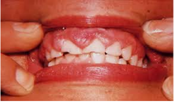
Results: A total of 48 (4%) patients were found to have reactive hyperplastic lesions. Focal fibrous hyperplasia is the most common type representing (91.7%). Peripheral giant cell granuloma was the second most common type representing (6.3%). The most risk factors were qat chewing, teeth grinding, cheek biting and poor oral hygiene.
Conclusion: In the current study, irritant fibroma and pyogenic granuloma were the most and least common oral reactive hypertrophic lesions, respectively. Compared with males, the prevalence of lesions in female patients was equal, and older age groups were more probable to grow oral reactive hypertrophic lesions. The most frequent location for lesions was the gingiva.
Peer Review History:
Received 5 April 2025; Reviewed 11 May 2025; Accepted 26 June; Available online 15 July 2025
Academic Editor: Dr. A.A. Mgbahurike , University of Port Harcourt, Nigeria, [email protected]
Reviewers:
Dr. George Zhu, Tehran University of Medical Sciences, Tehran, Iran, [email protected]
Dr. Heba M. Abd El-Azim, Damanhour University, Egypt, [email protected]
Downloads


Published
How to Cite
Issue
Section
Copyright (c) 2025 Universal Journal of Pharmaceutical Research

This work is licensed under a Creative Commons Attribution-NonCommercial 4.0 International License.


 .
.